Cutting the cement risk: practical steps to reduce residual cement around implant crowns
A new systematic review finds resin-containing cements leave the most undetected remnants; here’s a simple, low-cost protocol you can use on Monday to lower cement‑related peri‑implant problems.
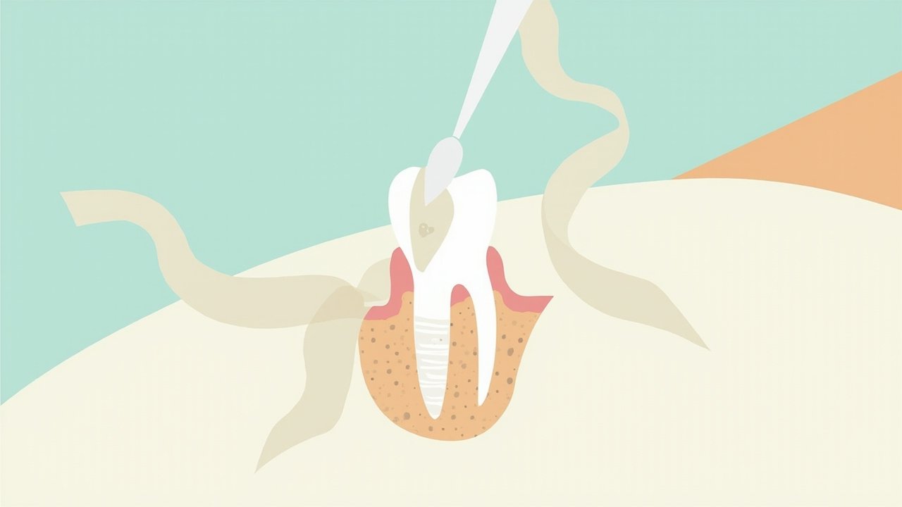
Residual luting cement remains a leading, preventable cause of peri‑implant mucositis and peri‑implantitis. A 2026 systematic review of the literature found that, across the studies available, cements that contain resin consistently left larger amounts of undetected subgingival residue, while zinc‑oxide based materials tended to leave the least. The review emphasises that most of the evidence comes from laboratory models, so clinical judgement remains essential. ([lifescience.net](
Clinical trials and recent RCTs back up the clinical relevance. In a randomized trial, nearly all cemented single crowns still showed detectable residual cement when assessed after cementation: 39 of 40 abutments (97.5%) had remaining material despite routine clean‑up. That shows how easy it is for small amounts to be missed and why a deliberate protocol matters. ([onlinelibrary.wiley.com](
Smaller amounts of residue matter. A recent intra‑patient clinical study measured the visible crown‑abutment surface area covered by remnants and found average values in the low single digits (around 1.5–2.0% of surface area for the materials tested). Even these small islands of cement can act as foci for biofilm and foreign‑body reaction if they sit submucosally. Choosing materials and techniques that reduce both the volume and the chance of submucosal placement is therefore high‑value prevention. ([pubmed.ncbi.nlm.nih.gov](
What to change in a modest private clinic (the Monday checklist). First, avoid definitive resin cements when margins will land subgingivally or where access is limited. Where you must use cement retention, prefer temporary or zinc‑oxide based systems in deep or interproximal sites, understanding their lower propensity for hidden residues. Second, keep the crown margin as shallow as the aesthetic result allows — studies show deeper margins correlate strongly with more undetected cement. Aim for no more than about 1 mm subgingival where feasible. ([lifescience.net](
Technique matters more than brand. Extra‑oral cementation on an analogue (pre‑seat the crown on a working model, remove gross excess, then seat in the mouth) and using PTFE tape or a small cotton pellet to block screw channels or venting holes reduce overflow and trapped cement. Multiple reviews and clinical investigations identify extra‑oral cementation and careful rehearsal with a replica abutment as the most effective manoeuvres to cut residual cement. If you must cement intra‑orally, use minimal cement volume and venting. ([pmc.ncbi.nlm.nih.gov](
Cleanup: tools and verification. Simple explorers and floss are often insufficient for subgingival traces. PEEK ultrasonic tips or dedicated plastic/PEEK scalers consistently remove more cement without damaging titanium surfaces and are affordable for most clinics. Use a periapical radiograph after cementation when your cement is radiopaque; when not radiopaque, rely on a careful visual‑tactile check and consider a short‑interval review at 1–2 weeks. ([pubmed.ncbi.nlm.nih.gov](
When to prefer screw‑retained. Where prosthetic geometry and occlusion permit, choose screw retention for posterior or non‑esthetic areas, or implants with limited access. Screw‑retained prostheses remove the whole problem of subgingival cement and simplify follow‑up. Remember, cement selection and technique are risk‑management, not absolute solutions; schedule recall and probe the peri‑implant sulcus regularly to catch early inflammation. ([onlinelibrary.wiley.com](
Limitations and pragmatic reading of the evidence. The 2026 systematic review drew mostly on in vitro work plus a small number of clinical studies; laboratory measurements of residue do not always translate one‑to‑one into peri‑implant disease. Still, the clinical trials and cohort data show real‑world risk: even small residues are common and removable with deliberate steps. These are low‑cost, high‑yield changes to your workflow that reduce avoidable complications.
Clinical takeaways
- If margins must be subgingival, avoid definitive resin cements — prefer temporary or zinc‑oxide‑based luting agents to reduce hidden remnants.
- Use extra‑oral cementation on an analogue, limit margin depth (keep <~1 mm subgingival when possible), and vent the crown to prevent trapping cement.
- Cleanup with PEEK/plastic ultrasonic tips and verify with radiograph when using radiopaque cement; schedule an early 1–2 week check to detect missed residue.
Sources
- Effect of luting materials on residual cement around cement-retained implant-supported dental restorations: A systematic review. International journal of oral implantology (Berlin, Germany) (2026)
- Residual Cement Excess of Composite Versus Glass Ionomer Cement in Implant‑Supported Single Crowns After Different Intraoral Exposure Times—A Randomized Controlled Clinical Trial. Clinical Oral Implants Research (2026). DOI: 10.1111/clr.70113
- An Intra-Patient Cohort Study on the Effect of Resin-Cement Types on Cement Remnants in Implant-Supported Restorations. The International Journal of Prosthodontics (2026). DOI: 10.11607/ijp.9757
- A Systematic Review of Cementation Techniques to Minimize Cement Excess in Cement-Retained Implant Restorations. International/PMC open review (2022)
- Effectiveness of PEEK ultrasonic tips and cement types on residual cement around implant-supported restorations. British Dental Journal (2025). DOI: 10.1038/s41415-025-8719-y